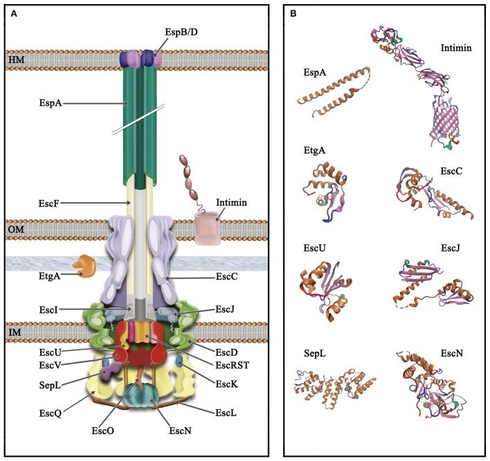
Figure 2.
Schematic representation of the type III secretion system of A/E pathogens. (A) The T3SS is divided into three main parts, from top to bottom (i) extracellular appendages: translocation pore (inserted into the host membrane, HM), filament and needle; (ii) basal body: consisting of three membrane rings that span the inner and outer membrane (IM and OM, respectively) connected through a periplasmic inner rod. The IM rings house the export apparatus components; (iii) cytoplasmic components: the C-ring, the ATPase complex and the gatekeeper protein. The outer membrane protein intimin and the PG lytic enzyme EtgA are also illustrated. (B) Solved protein structures of the depicted T3SS components. Protein Data Bank (PDB) accession numbers: SepL, 5C9E; EscN, 2OBM; cytoplasmic C-terminal domain of EscU, 3BZL; periplasmic domain of EscC, 3GR5; EtgA, 4XP8; periplasmic domain of EscJ, 1YJ7; the EspA structure was obtained from that of the CesAB/EspA complex, 1XOU, chain A; transmembrane beta-domain of intimin, 4E1S and its C-terminal domain, 1F00. Protein structures are displayed as ribbon diagrams and were colored according to their secondary structure.

