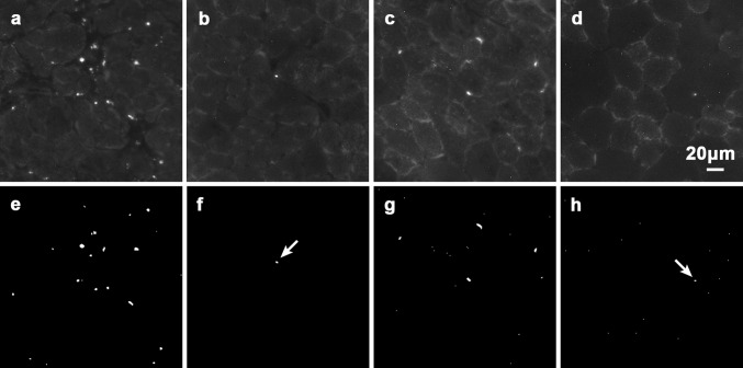
Fig. 2.
Typical raw images of autofluorecent LF granules in 7-μm-thick sections of diaphragm muscles of a 8-week-old mdx mice and b WT normal mice and EDL muscles of c mdx mice and d WT normal mice. Their corresponding binary images are shown in (e–h). As a measure of the granule sizes, two extracted LF granules (arrows) are shown in (f, h): the granule areas are 5.07 and 3.04 μm2, respectively, and their Feret diameters 3.56 and 2.57 μm, respectively

