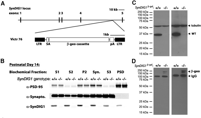
Figure 2.
Characterization of SynDIG1 β-gal mutant mice. A, Schematic of mouse SynDIG1 locus showing the insertion site of the β-geo cassette between exon 4 and exon 5 to create SynDIG1 β-gal homozygous mutant mice. SynDIG1 expression is disrupted and replaced with β-gal expression. SA, Splice acceptor; pA, synthetic polyA signal/transcriptional blocker; LTR, viral long-terminal repeat segment. B, Immunoblots of biochemical fractions from P14 wild-type (+/+) and SynDIG1 β-gal homozygous mutant (−/−) mice using a LI-COR system. Fractions loaded are S1-, S2-, P2-, Syn-, S3-, and PSD-enriched fractions. As a control for the biochemical fractionation, PSD-95 is enriched in the PSD fraction and de-enriched in S2 and S3 while synaptophysin (Synapto) is absent from PSD and enriched in S3. Note that (+) refers to the unmodified SynDIG1 allele. C, Chemiluminescence immunoblot to detect SynDIG1 expression in brain lysates from wild-type (+/+) and SynDIG1 β-gal homozygous mutant (−/−) mice. Arrowheads indicate full-length protein and tubulin (loading control) at 2 min (left) and 4 h (right) exposure times. Note that (+) refers to the unmodified SynDIG1 allele. D, Chemiluminescence immunoblot to detect β-geo reactivity in brain lysates from WT (+/+) and homozygous (−/−) mutant mice. Arrowheads indicate bands for β-geo and mouse IgG (recognized by secondary antibody) at 2 min (left) and 4 h (right) exposure times. Note that (+) refers to the unmodified SynDIG1 allele.

