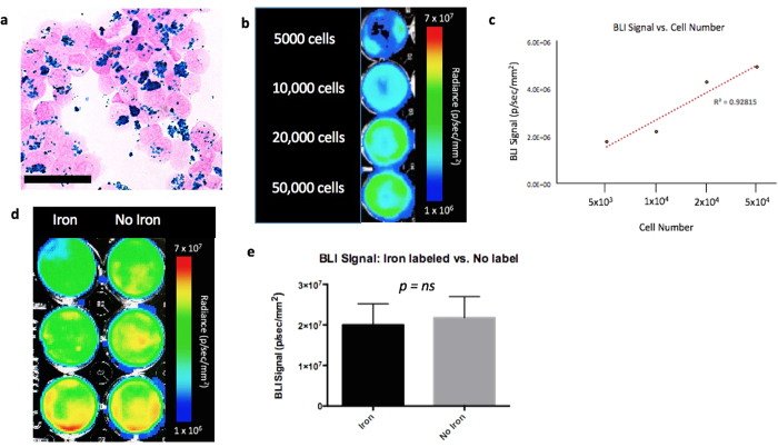
Figure 2.
In Vitro Experiments (a) Perl’s Prussian blue stain identifies iron labelled cells in blue (scale bar x 100). (b) JIMT1BR3-Fluc/GFP + cells seeded at various concentrations (c) A strong linear correlation is seen between cell number and BLI signal; R2 = 0.928 (d) MPIO labeled JIMT1BR3-Fluc/GFP + cells (L) and non labeled JIMT1BR3-Fluc/GFP + cells (R). (e) There was no significant difference in BLI signal detected in cells that were labeled with MPIO and cells that were not labeled (ns, p value = 0.82). These results are representative of three experiments with three wells of each condition.

