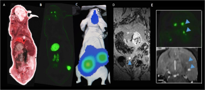
Figure 7.
Cryoviz Imaging: (A) Brightfield image of cryoviz mouse imaged on day 28 (B) Fluorescence microscopy detects GFP + brain tumours and body tumours in same slice as brightfield image (C) Corresponding day 28 BLI and (D) Whole body MRI (E) Brain metastases (blue arrows) in cryo-image and corresponding MRI slice in the same orientation. Scale bar = 0.50 mm.

