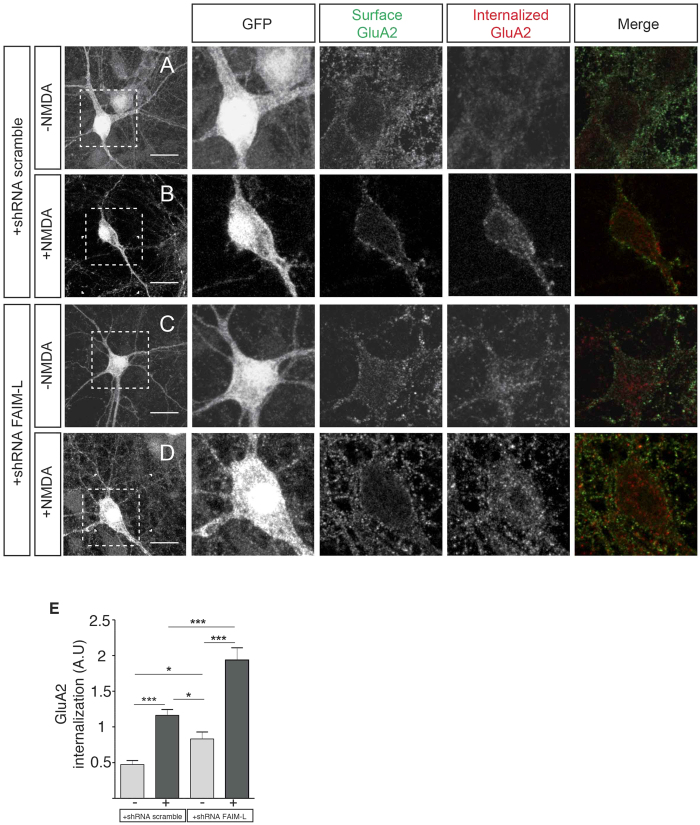
Figure 3. FAIM-L actively participates in GluA2 internalization induced by chemical LTD.
(A–D) antibody-feeding internalization assay for endogenous GluA2 in hippocampal neurons stimulated with NMDA (50 μM for 15 min). Neurons were infected with lentiviruses containing the shRNA constructs (scrambled or FAIM-L). The figure shows triple-label immunostaining for infected GFP-positive neurons (first and second column), surface-remaining GluA2 (third column, green in merge), internalized GluA2 (fourth column, red in merge), and merge (fifth column). Individual channels are shown in grayscale. Images in columns 2, 3, 4 and 5 represent magnifications from selected areas of the first columns. The scale bar represents 20 μm. (E) quantification of GluA2 internalization in the indicated conditions calculated as in Fig. 2(I). Results were not normalized to untreated cells (−NMDA). As a consequence, A.U have no dimensions. N = 48 to 53 neurons from 3 independent experiments, for each group. Data represent means ± SEM and were analyzed by the One-way ANOVA test followed by Newman-Keuls multiple comparison post-hoc test, ***p < 0.001, *p < 0.05.

