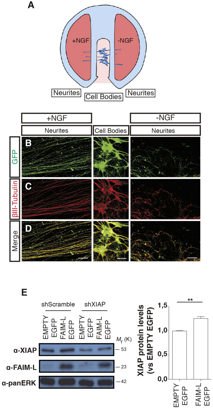
Figure 6. FAIM-L stabilizes XIAP protein levels in DRG neurons.
(A) Schematic representation of a Campenot chamber. Cell bodies and axonal compartments were subjected (−NGF) or not (+NGF) to NGF withdrawal for 24 h. (B–D) panels are representative confocal images of the DRG neurons cultured in a Campenot chamber. (E) DRGs neurons were infected with either EMPTY-EGFP or FAIM-L-EGFP vectors. Protein levels of XIAP and FAIM-L were determined by Western blot 48 h after infection with overexpressing and silencing vectors. In the scrambled shRNA condition, the intensity of the bands relative to the control (“EMPTY EGFP”) was quantified using ImageJ software. Data are represented as the mean ± SEM of three independent experiments. Student’s t-test was used to calculate significant levels between the indicated groups. **p < 0.01. Scale bar 50 μm.

