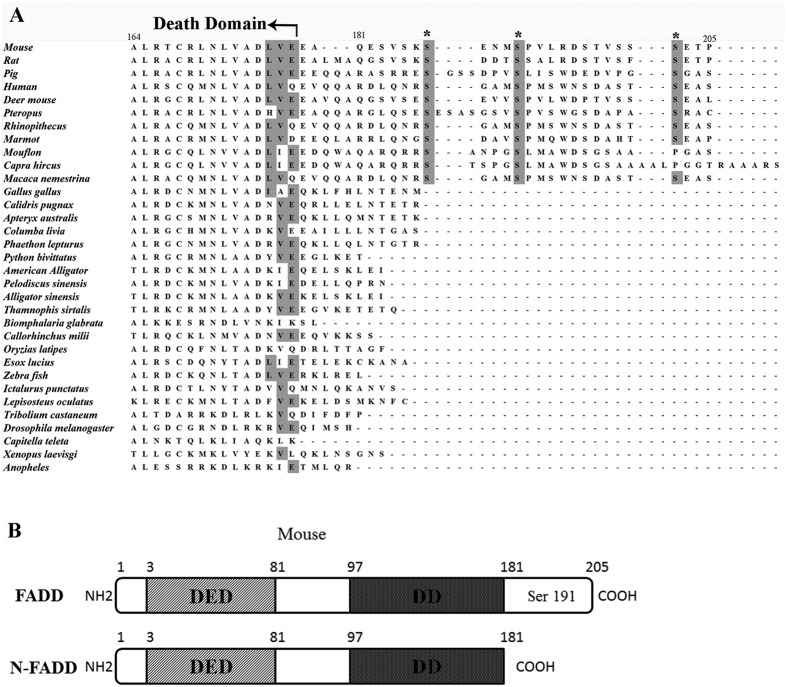
Figure 1. Alignment of FADD proteins from different species and diagram of N-FADD.
(A) Alignment of FADD proteins from several different species. Only the C-terminal regions are shown to highlight the conservation of the known phosphorylation sites, ser 191 of mouse FADD (equivalent to Ser194 of human FADD), and the other two putative ones, Ser 187, Ser 202, are indicated by asterisks to highlight their conservation in mammals. The residue numbers are based on the corresponding amino acid residue position in the mouse FADD protein. All of the FADD protein sequences were obtained from NCBI and aligned by Clustalw. (B) FADD and N-FADD of mouse. The numbers indicate the amino acid residue position in the mouse FADD protein.

