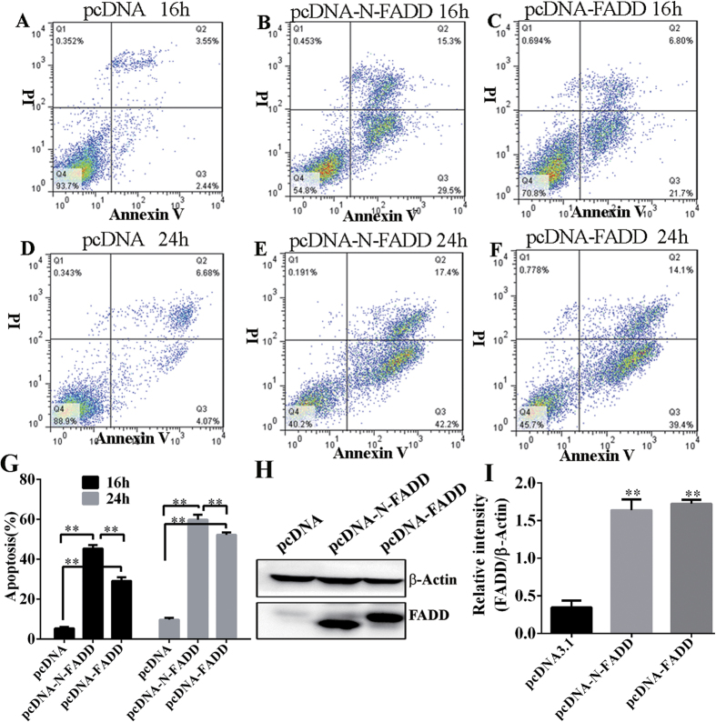
Figure 2. Overexpression of FADD or N-FADD induced apoptosis of B16F10 melanoma cells in vitro.
(A–C) Representative FACS analysis of Annexin V and propidium iodide (PI) staining after transfection of FADD or N-FADD expressing or empty vectors for 16 h. (D–F) Representative FACS analysis of Annexin V and PI staining after transfection of FADD or N-FADD expressing or empty vectors for 24 h. (G) Overexpression of FADD or N-FADD induced apoptosis of B16F10 melanoma cells (Mean ± SD, n = 3 independent experiments); **P < 0.01. (H) Western blots analysis of expression of FADD after transfection of FADD or N-FADD expressing or empty plasmids in B16F10 cells. β-actin was served as loading control. (I) Statistical analysis of western blot by Image J; **P < 0.01, compared with pcDNA3.1. One representative of three independent experiments is displayed.

