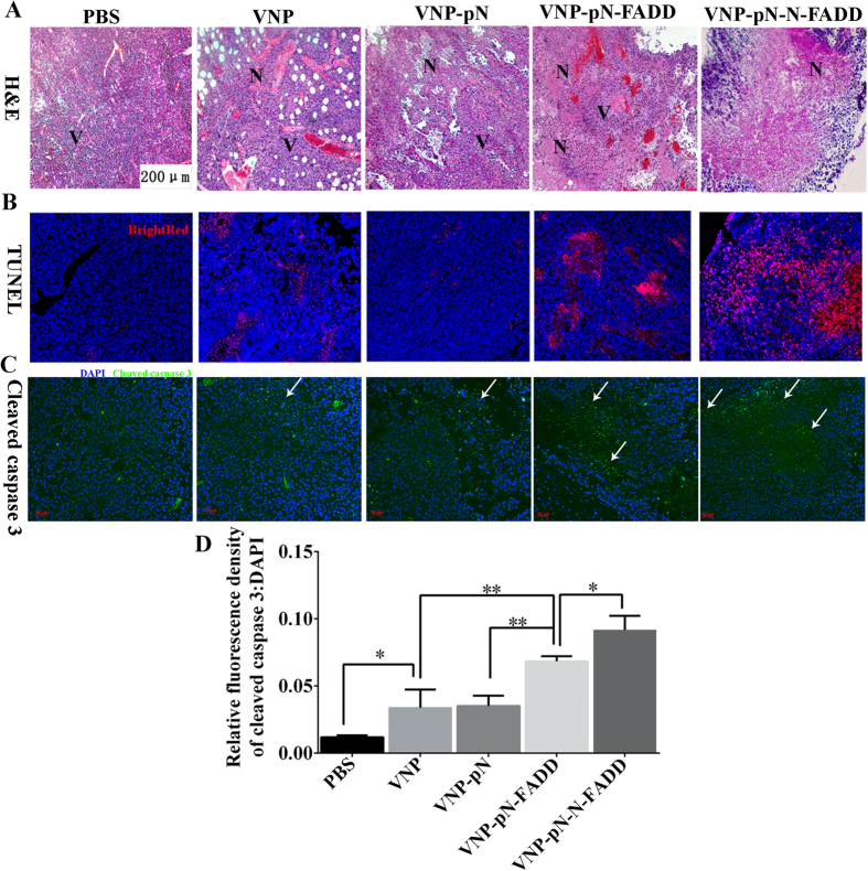
Figure 6. Salmonella typhimurium strain VNP20009 carrying FADD or N-FADD induced apoptosis of melanoma cells.
(A) H&E staining of the tumor sections. The representative images (100×) revealing necrotic areas of B16F10 tumor tissue treated with PBS, VNP, VNP-pN, VNP-pN-FADD and VNP-pN-N-FADD; N, necrotic tumor regions; V, vital tumor regions. (B) TUNEL assay of melanoma. The representative images (100×) revealing apoptosis of tumor cells treated with appropriate VNP strains; bright red, apoptotic cells. (C) Immunofluorescence staining of cleaved caspase-3 (stained green and indicated with blank arrows), the representative images (200×) revealing cleaved caspase-3 in B16F10 tumor tissue treated with PBS, VNP, VNP-pN, VNP-pN-FADD and VNP-pN-N-FADD. (D) Quantification analysis of relative cleaved caspase-3 expression level (fluorescence intensity of cleaved caspase-3 DAPI) by Image J software, *P < 0.05, **P < 0.01, bar represents the mean ± SD of five optical fields. Each experiment was carried out in triplicate.

