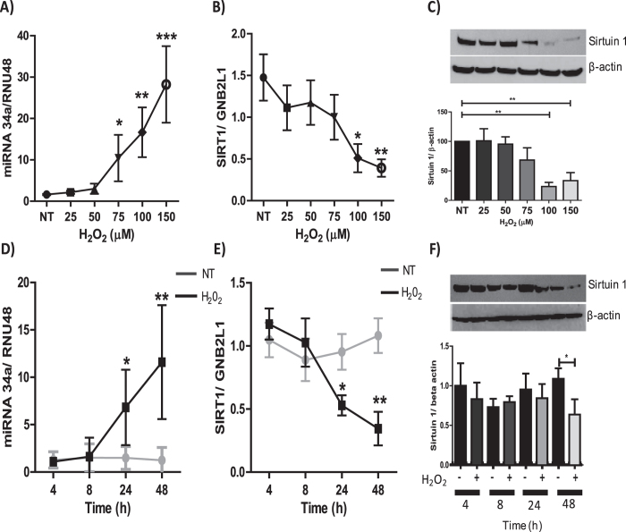
Figure 2. Correlation between oxidative stress-mediated reduction in SIRT1 and increased miR-34a expression.
BEAS2B cells were stimulated for 48 hours with H2O2 at concentrations of 25, 50, 75, 100 and 150 μM, and protein or RNA extracted. (A) RNA was extracted to examine miR-34a (n = 6) (B) and SIRT1 (n = 6). (C) Protein was extracted and SIRT1 protein expression was determined by SDS-PAGE/Western blotting normalized to β-actin (n = 5). BEAS2B cells were stimulated for 4, 8, 24 and 48 hours with 100 μM H2O2 and protein and RNA extracted, (D) changes in miR-34a expression was examined (n = 5), as well as changes in (E, F) SIRT1 gene and protein expression (n = 5). The band density of each blot is represented as a histogram and and is the average of all experiments performed. Data are means ± SEM, analyzed by Kruskal–Wallis test with post hoc Dunns and One-way Anova with post hoc Bonferroni *P < 0.05, **P < 0.01, ***P < 0.001.

