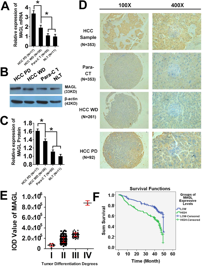
Figure 1. MAGL levels in tissue specimens and clinical relevance.
MAGL mRNA levels (A) and protein levels (B,C) were examined by RT-PCR and western blot. Both mRNA levels and protein levels increased as the malignancy increased [from normal liver tissue (NLT) and para-carcinoma tissue (Para-CT) to HCC tissue well-differentiated (HCC WD) and HCC tissue poor differentiated (HCC PD)] in tissue specimens from HCC patients (*p < 0.05). These results were confirmed with IHC of TMA (D, 100× magnification; 400× magnification). MAGL protein was stained brown by IHC in the tissue slices, and stain was deeper when malignance increased in liver and tumor tissues. Patients were distributed according to MAGL protein level measured by IOD value by Image-Pro Plus 6.0 software (E). MAGL protein levels increased significantly with decreasing of tumor differentiation degrees15 of HCC (comparison among groups, p < 0.05). Kaplan Meier method for survival analysis (F) also demonstrated the MAGL low-expression group (MAGL IOD value <200000) showed significantly better survival than the MAGL high-expression group (MAGL IOD value >200000, p < 0.05).

