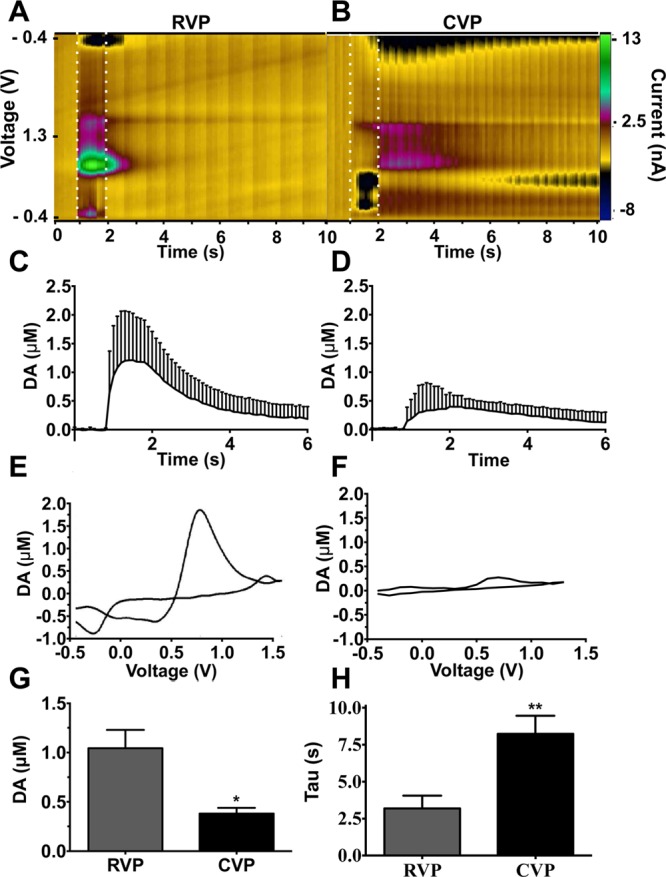
Figure 5.

Dopamine release in RVP and CVP. Stimulation of RVP produced significantly greater dopamine release compared with stimulation of CVP (A–G, 1.04 μM vs 0.38 μM, respectively; p = 0.011, RVP n = 19, CVP, n = 12, two-tailed t test). Dopamine clearance is slower in CVP than RVP (H, p = 0.002, RVP n = 19, CVP n = 12, two-tailed t test). Representative color plots (A, B) and voltammagrams (E, F) are shown. Current time traces (C, D) are cumulative. Dotted lines indicate stimulation boundaries.
