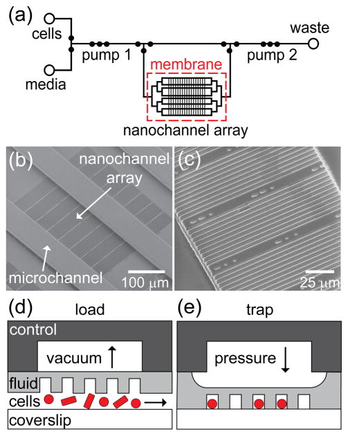
Figure 1.
(a) Schematic of the fluid layer of the microfluidic device with an integrated nanochannel array for monitoring growth and development of single cells over multiple generations. Open circles represent reservoirs, and closed circles represent valves. Three valves in series are used as peristaltic pumps and are labeled pumps 1 and 2. A large open region in the control layer above the nanochannel array (~4 mm × 3 mm) actuates the cell trapping region. (b) Scanning electron microscope (SEM) image of a section of the SU-8 fluid-layer master that shows 80 nanochannels with widths from 600 to 1000 nm positioned orthogonal to the microchannels, which deliver nutrients to the nanochannels. This set of 80 nanochannels is one of 16 sets of channels arranged in a 4 × 4 grid. (c) SEM image of a subset of nanochannels replicated in poly(dimethylsiloxane) (PDMS). Schematic of pneumatic actuation with (d) vacuum applied to raise the nanochannel array and load cells and (e) pressure applied to lower the nanochannel array and trap cells for growth and analysis. The control layer, fluid layer, and coverslip constitute the three layers of the device.

