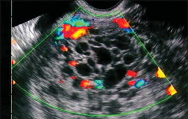
Figure 1.

Transvaginal ultrasound in a patient with bleeding at 14 weeks of pregnancy, showing an enlarged uterus with an endometrial cavity filled with amorphous material with multiple anechoic areas, suggestive of complete hydatidiform mole. Note the absence of embryonic tissue and its attachments. Note that, in the Doppler flow study, there was no vascular flow among the vesicles, indicating their avascular nature.
