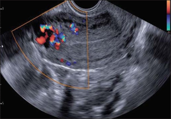
Figure 6.

Transvaginal ultrasound, acquired during post-molar pregnancy followup, when hCG levels were elevated. Note the presence of hypoechoic areas in the myometrium, resembling the hypervascular “Swiss cheese” aspect, suggestive of an invasive mole, on the Doppler flow study.
