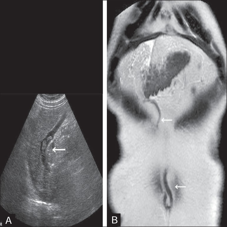
Figure 7.
A: Longitudinal view from B-mode ultrasound showing the recanalized paraumbilical vein partially filled with hypoechoic material, forming a partial thrombus B: T2-weighted magnetic resonance imaging, in the coronal plane, showing the entire trajectory of the paraumbilical vein, from its origin at the round ligament to the umbilicus (arrow).

