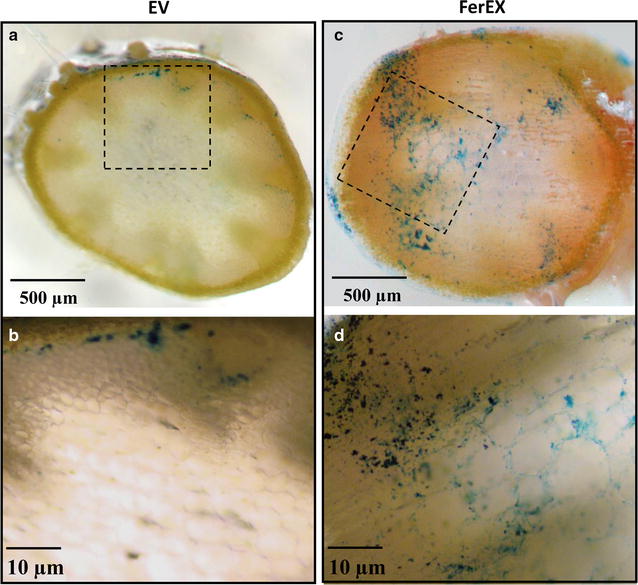
Fig. 3.

Perls’ Prussian blue staining of cross sections from fresh stem tissues in transgenic Arabidopsis plants expressing ferritin that secreted extracellularly. Brightfield optical microscopy showing Perls’ Prussian blue staining of empty vector (EV) control (a, b), and extracellular ferritin-expressing transgenic plant shoot tissue (FerEX) (c, d). b, d are images taken at a higher magnification of the black boxes outlined in a and c, respectively
