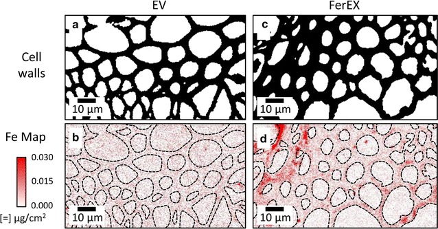
Fig. 4.

X-ray fluorescence microscopy (XFM) elemental maps for Fe in stem cross sections of extracellular ferritin-expressing and control Arabidopsis plants. The 2-micron-thick cross sections were cut from senesced stems from empty vector (EV) control (a, b) and extracellular ferritin-expressing (FerEX) (c, d) plants. Cell-wall images (a, c) were constructed from binary images of potassium XFM maps. The dashed lines were drawn from the cell-wall images and overlayed on the iron maps (b, d) to more easily distinguish iron intensity inside the cell walls. The intensities in both iron maps (b, d) were scaled the same, and the iron is observed in the FerEx cell walls (d) by noting that the iron intensity in the cell walls is higher than the background iron intensity observed in the empty cell lumina
