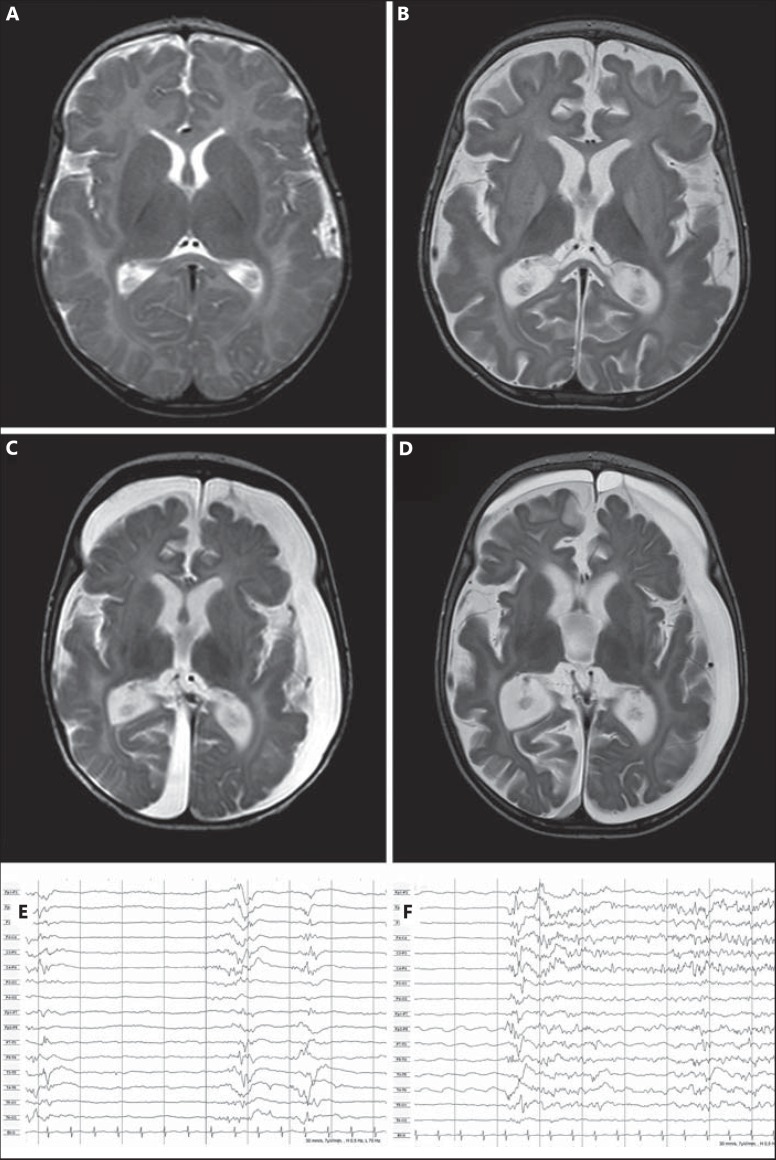
Fig. 2.
Brain MRI of the IP with OS. Axial T2-weighted images at seizure onset at an age of 3 months (A) and at 4 (B), 5 (C) and at an age of 6 months (D), respectively, demonstrating progressive supratentorial brain atrophy. EEG at 14 weeks displaying burst-suppression pattern (E) and onset of a tonic seizure (F). Both are the characteristic features of OS.

