
Fig. 1.
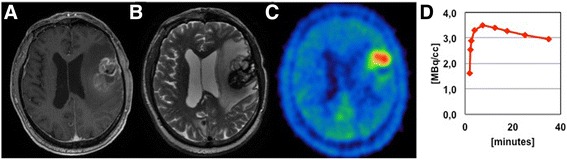
MRI and 18F-FET PET findings in a recurrent brain metastasis after focused high dose radiotherapy (Patient nr. 30). A ring-like contrast enhancement in the T1-weighted images (a), as well as an extensive edema in the T2-weighted images is seen (b). Both a focal 18F-FET uptake (TBRmax 3.5, TBRmean 2.3) and decreasing TACs can be observed in static and dynamic PET analysis (c, d)