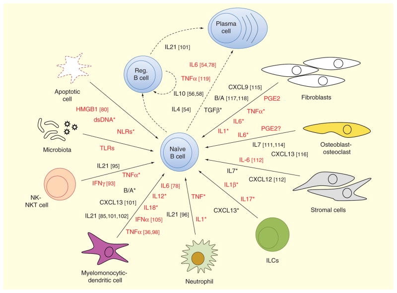
Figure 1. Innate cell-mediated inflammatory signals influencing B-cell function.
A naive B cell can be influenced by many signals from their immediate environment to acquire a plasmablast/plasma cell or regulatory B-cell fate. Different cells can contribute to the milieu of signaling molecules at a given time. Cell types considered in this graph include either cells that are structurally inherent to a certain tissue (osteoblasts, fibroblasts, stromal cells) or cells of the innate immune system (neutrophils, myelomonocytic cells, DCs, ILCs, MDSCs, NK and NKT cells). Due to their potential contribution in an inflammatory environment, we have included molecules derived from apoptotic cells and the local microbiota. Despite their profound differences, for simplification purposes, myelomonocytic and dendritic cells, as well as NK and NKT cells, have been grouped. For the same reason, we have also omitted differentiating fDCs versus nonfollicular DCs or including MDSCs. Molecules labeled in red indicate a strong proinflammatory bias, while black ones represent molecules implicated in recruitment, survival and proliferation of cells or suppressors of inflammation. Molecules indicated with an asterisk are well described in previous reviews like [3,5,98]. For the purpose of clarity, not all cell types and mediator molecules mentioned in the text are depicted in the figure.
B/A: BAFF/APRIL; DC: Dendritic cells; HMGB1: High-mobility group box 1 protein; ILC: Innate lymphoid cell; MDSC: Myeloid-derived suppressor cell; NK: Natural-killer; NLR: NOD-like receptors; PGE2: Prostaglandin E2.

