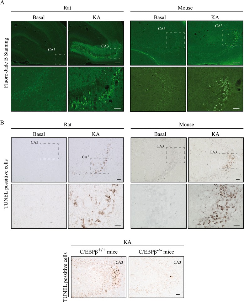
Fig. 1.

Excitotoxic brain injury induced by KA injection in the hippocampus. Animals were injected in the right hemisphere with KA and sacrificed 72 h post-injection. a Representative images of Fluoro-Jade B staining in the CA3 region of the hippocampus. Scale bars, 200 μm (rat) and 250 μm (mouse). Inset scale bars, 50 μm (rat) and 25 μm (mouse). b Representative images of TUNEL staining in the CA3 region of the hippocampus. Scale bar, 50 μm. Inset scale bar, 25 μm
