
Figure 5.
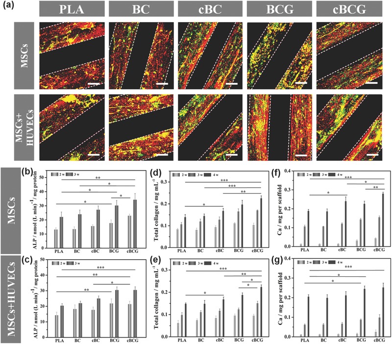
a) Immunofluorescence staining of the vascularized bone formation in the dynamic co‐culture condition. The fluorescence images for anti‐vWF (green) and OPN (red) showed that the cBCG scaffold possessed more vascular‐like network and osteogenesis than other control groups. The scale bars indicate 100 μm. b,c) Quantification of ALP activity. d,e) Total collagen synthesis. f,g) Quantification of calcium deposition content on different scaffolds comparing the dynamic co‐culture with dynamic monoculture. All data showed that the cBCG scaffold enhanced the osteogenic differentiation.