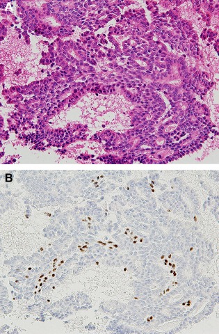
Figure 6.

Intraductal papilloma diagnosed by a returned cell block (Case 2). (A) Note the papillary arrangement of epithelial cells with mild cellular atypia admixed with myoepithelial cells (HE stain, ×200); (B) The myoepithelial cells are confirmed by p63 immunostaining (×200). [Color figure can be viewed in the online issue, which is available at wileyonlinelibrary.com.]
