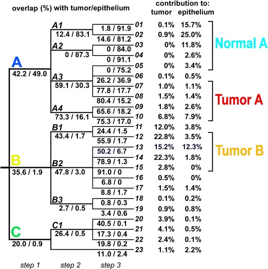
Figure 3.

Description of clusters detected in three steps of concomitant unsupervised clustering of all tissue preparations. Shown is an overlap between each cluster and regions corresponding to tumor and normal epithelium. Contribution of each cluster detected in the third step of segmentation (clusters 01 to 23) to expert‐defined areas is presented on the right.
