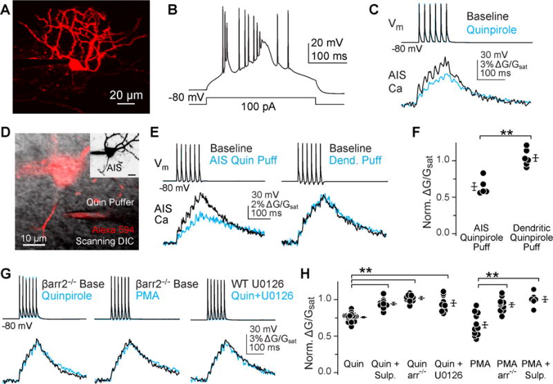
Fig 1. D3R-dependent regulation of AIS Ca is β-arrestin dependent.

A) 2-photon z-stack of cartwheel cell, visualized with Alexa 594.
B) Characteristic burst spiking of cartwheel cells evoked with somatic current injection.
C) Quinpirole (2 μM) suppresses spike-evoked Ca influx in the AIS, as seen previously (Bender et al., 2010).
D) Simultaneous 2P fluorescence and scanning-DIC was used to position a puffer pipette near the AIS. Inset: maximum intensity z-stack of neuron, highlighting that the AIS was well separated from dendrites.
E) AIS Ca modulation occurred only when quinpirole was applied near the AIS. Somatic voltage and AIS Ca transients are shown before (black) and following (cyan) local application of quinpirole.
F) Summary of local quinpirole application across cells. Circles are single cells, bars are mean ± SEM. Data are normalized to pre-drug conditions within each experiment. ** p < 0.01, Mann-Whitney.
G) AIS Ca modulation was not observed in β-arrestin-2 knockouts, or when blocking ERK1/2 signaling with U0126. Drugs were applied to ACSF.
H) Summary of β-arrestin-dependent signaling in the AIS. Format as in panel f, ** p < 0.01, ANOVA, Newman-Keuls. Sulpiride is abbreviated “sulp.”
