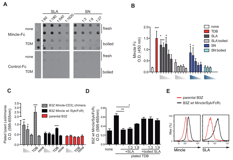
Figure 1. Leishmania releases a soluble ligand for Mincle.
(A) Dot blots for Mincle-Fc (top) or control-Fc (bottom) with membranes spotted with fresh or boiled soluble Leishmania extracts (SLA) (left) or supernatants (SN) (right) from the indicated dilutions of stationary cultured parasites. Culture medium (none) or TDM were used as controls. (B) ELISA with Mincle-Fc of different doses of SLA or supernatants (fresh or boiled) from L. major promastigotes and controls (none or TDB). (C) NFAT reporter activity in response to 106, 105 or 104 of plated lysed-Leishmania or TDB in B3Z cells expressing human Mincle-CD3ζ chimera, WT mouse Mincle receptor co-expressing Syk and FcRγ, or the parental cells. (D) NFAT reporter activity in B3Z cells expressing WT mouse Mincle receptor, FcRγ, and Syk and exposed to plated TDB in the presence of the indicated dilutions of fresh or boiled SLA. (E) Staining with anti-Mincle (left) and fluorochrome-labeled SLA on control and Mincle-expressing. (A, E) Data are from one representative experiment of four (A) or three (E) performed. (B, C, D) Bars show arithmetic mean + SEM corresponding to three independent experiments. * p < 0.05; ** p < 0.01; *** p < 0.001 (one way ANOVA with Bonferroni post-hoc test).

