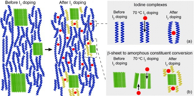
Figure 12.

Schematic of neat and I2-doped spider silk, and proposed I2-silk interaction mechanisms. Left: general spider fibroin structure before I2 doping showing β-sheet crystallites (green rectangles) connected by amorphous chains composed of 31 helices (blue coils). Right: spider fibroin structure after I2 doping showing (a) iodine (red dots) complexes between the amorphous chains, and (b) conversion of some part of β-sheet into additional amorphous constituents (yellow coils).
