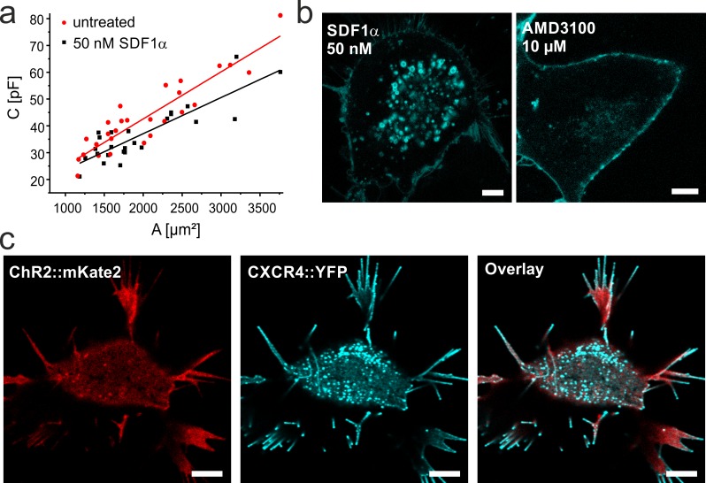
Fig 2. SDF1-mediated CXCR4-internalization in NG108-15 cells.
a. Plot of cell capacitance (including stray capacitance) measured in whole cell configuration by patch-clamp technique against the membrane area. Cells were either not treated (red) or treated with 50 nM SDF1α (black) for >40 min. The membrane area was calculated from the measured cell diameter assuming a spherical shape of the cell. The specific cell capacitance (slope of the linear fit) decreases in presence of SDF1α. b. Confocal laser scanning micrographs of NG108-15 cells expressing CXCR4::eYFP in the presence of the inhibitor AMD3100 or the agonist SDF1α as indicated. While strong internalization is observed with SDF1α, only few vesicles are observed in presence of the inhibitor. c. Co-expression of CatCh::mKateA (red) and CXCR4::eYFP (cyan) in presence of 50 nM SDF1α. Note that after binding of SDF1α CXCR4 is internalized while CatCh mainly remains in the plasma membrane and is not directly affected by this growth factor (S3 Fig). Scale bars represent 5 μm.

