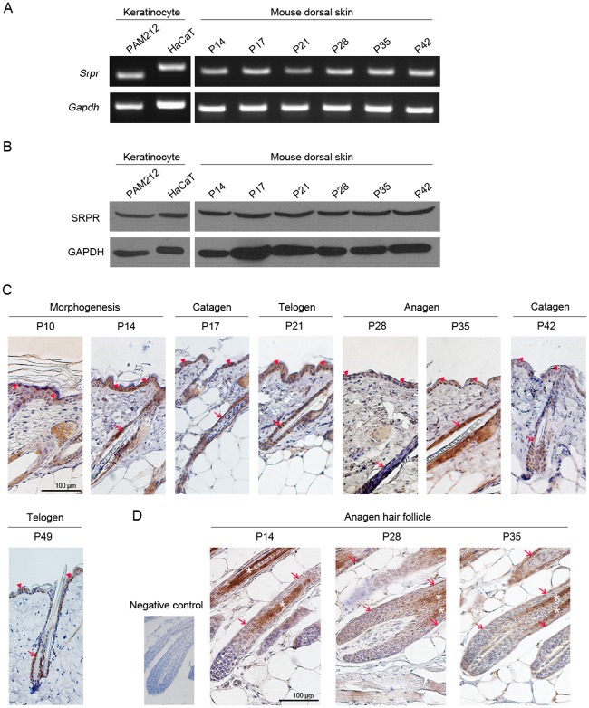
Fig 1. Abundant expression of SRPR in mouse epidermal keratinocyte.
(A-B) Srpr was abundantly expressed in keratinocyte (PAM212 and HaCaT cell) and mouse dorsal skin at both mRNA by RT-PCR (A) and protein level by western blot analysis (B). (C-D) SRPR expression in dorsal skin (C) and HF (D) of BALB/C mice at postnatal days P10, P14, P17, P21, P28, P35, P42 and P49 by immunohistochemistry. Brown signals indicated the SRPR-positive epidermis cells (arrowhead), HF (arrow) and hair cortex (star). Scale bar = 100 μm.

