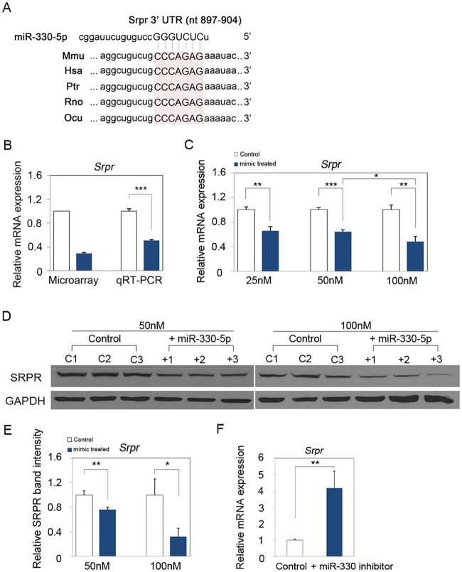
Fig 3. MiR-330-5p regulates Srpr expression in mouse epidermal keratinocyte.
(A) Targetscan algorithm predicted that a conserved binding sequence of miR-330-5p was present in the 3’ UTR of Srpr mRNA. (B) Real-time quantitative PCR was performed to validate the result of previous study using the same total RNA used for microarray analysis. (C) MiR-330-5p down-regulated the Srpr expression at 25nM, 50nM and 100nM mimic transfected PAM212 cells. The data was normalized against Gapdh expression. (D) Western blot analysis also revealed that SRPR protein level was decreased at 50nM and 100nM miR-330-5p mimic transfected PAM212 cells in comparison to the control. β-actin was used as a loading control. (E) Quantitative analysis of western blot using ImageJ software. The SRPR band intensity was normalized against GAPDH expression. (F) Srpr expression was significantly increased by inhibition of miR-330-5p. (B-F) Results are the average of three independent experiments. *P<0.05; **P<0.01; ***P<0.001.

