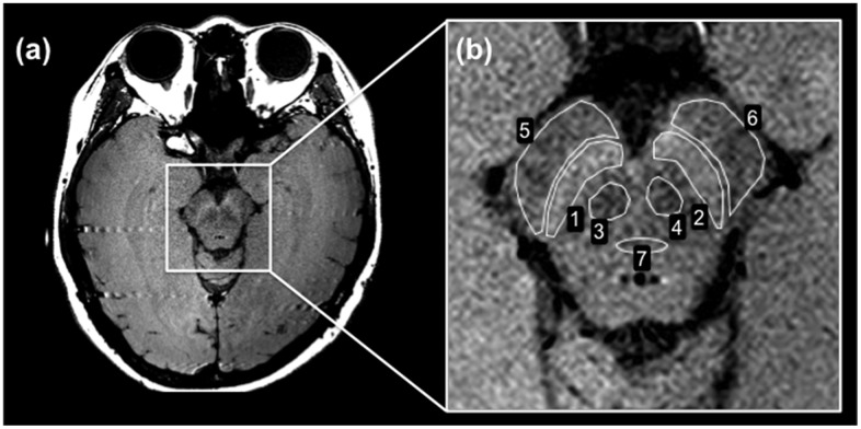
Fig 1. Neuromelanin-sensitive MRI of the lower midbrain.
(a) 2D-neuromelanin-sensitive MRI. The SNc appears as the high SI area. The SCP is the small low SI area below the SNc. The CP is above the SNc. The MT is located in the middle of the lower midbrain. (b) The ROIs for measuring the SI were traced manually at the following locations on a single slice: SNc (ROIs 1 and 2), SCP (ROIs 3 and 4), CP (ROIs 5 and 6), and MT (ROI 7).

