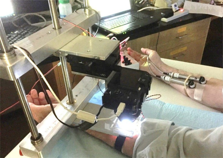
Fig 1. Sweat bubble imaging and evaporimetry.
Here the left arm is being tested using sweat bubble imaging and the right with evaporimetry. The reservoir slides into the holder which is held in fixed position to the frame; the camera can be precisely positioned using micrometer drives. Two probes are used for evaporimetry: one stays on the same patch of skin throughout and serves as a control; the other is removed to allow for injections and then replaced. Two tattooed sites on each arm were used to ensure that the same populations of identified glands were sampled by each assay.

