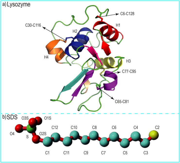
Fig 1. The structures of human lysozyme and sodium dodecyl sulfate.
(a) The native structure of human lysozyme represented as a new cartoon model. The α- helix structures are shown with the letter H and C6-C128, C30-C116, C65-C81, and C77-C95 represent disulfide bounds in the human lysozyme structure. (b) The structure of an SDS surfactant molecule with its polar head group (in red and green) and hydrophobic tail (in cyan and yellow) is shown as a ball and stick model.

