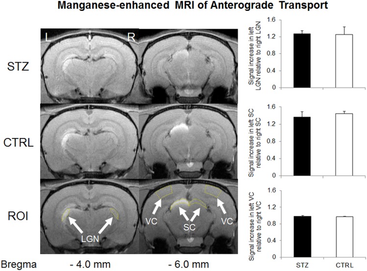
Fig 6. Manganese (Mn)-enhanced MRI of anterograde Mn transport along the visual pathway in streptozotocin (STZ) and sham control (CTRL) groups.
(Left panel) Mn-enhanced MRI of the visual brain nuclei at 1 month after systemic STZ or CTRL administration, and 1 day after intravitreal MnCl2 injection into the right eye. The regions of interest (ROI) for quantitative measurements were illustrated on both sides of the lateral geniculate nucleus (LGN), superior colliculus (SC) and visual cortex (VC) in yellow (arrows). Note the signal enhancements in the left lateral geniculate nucleus and left superior colliculus of both groups; (Right panel) Quantitative comparisons of Mn signal enhancements in the visual brain nuclei. Significantly higher signal intensities were found in the left LGN and SC than the right ones in both STZ and CTRL groups (Post-hoc Sidak’s multiple comparisons correction tests, p<0.01). No apparent difference in signal intensity was found between left and right VC in either group (Post-hoc Sidak’s multiple comparisons correction tests, p>0.05). No apparent difference in Mn enhancement was found in the lateral geniculate nucleus, the superior colliculus or the visual cortex between the two groups (Post-hoc Sidak’s multiple comparisons correction tests, p>0.05).

