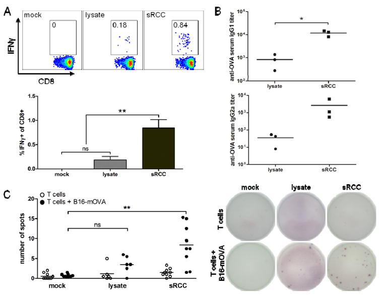
Figure 7.
Cellular and humoral response to sRCC vaccinations in naive mice. (A)–(B) Cellular and humoral response to vaccination with sRCCs or cell lysate derived from B16-WT cells, and loaded or admixed, respectively, with exogenous OVA protein. (A) Representative flow cytometry plots with mean frequency of IFNγ-positive cells among live CD8+ cells in peripheral blood (top), and quantification of data (bottom). Cells were stimulated in vitro with peptide. (B) anti-OVA IgG1 and IgG2a serum titers, on day 8 post-vaccination. Naive mice were vaccinated subcutaneously with either 370 μg B16-WT sRCCs loaded with 70 μg OVA and 90 ng MPLA, or cell lysate derived from the same number of B16-WT cells (5e6) and admixed with the same amount of OVA and MPLA. (C) Cellular response to vaccination with sRCCs or cell lysate derived from B16-mOVA cells. ELISPOT analysis of IL-2-producing T cells from spleens of mice following overnight co-culture with parent B16-mOVA cells on day 4 post-boost (day 22 post-vaccination). Naive mice were vaccinated subcutaneously on days 0 and 2 (prime), and days 16 and 18 (boost), with either 370 μg B16-mOVA sRCCs loaded with 18 μg CpG and 180 ng MPLA, or cell lysate derived from the same number of B16-mOVA cells (5e6) and admixed with the same amount of CpG and MPLA. Quantification of spots (left) and representative images of ELISPOT membranes (right). Values represent the mean ± SD, *p<0.05, **p<0.001. Data in (C) analyzed using one-way ANOVA, followed by Tukey post-test.

