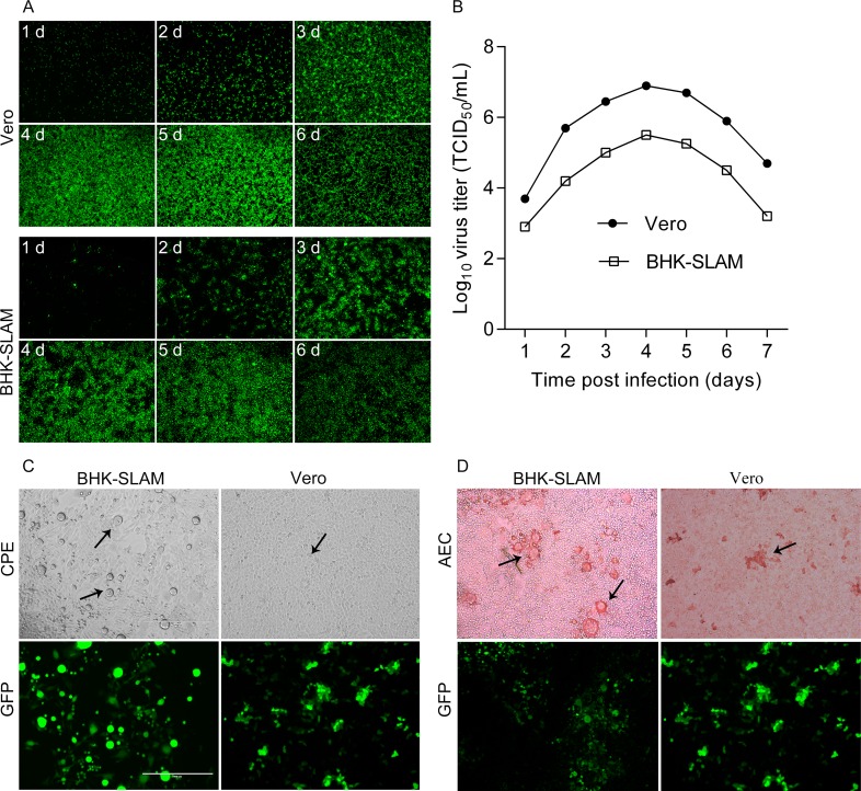
Fig 3. Comparison of BHK-SLAM cells and Vero cells.
(A) BHK-SLAM cells and Vero cells were grown to 80%–90% confluence in 24-well plates. Then, they were infected with rPPRV/GFP at an MOI of 0.01. At various time points after infection (every 24 h), images of GFP expression were taken. (B) Viruses at different time points were collected and stored at −70°C, and viral titers (TCID50) were determined in Vero cells. (C) BHK-SLAM and Vero cells were fixed at the same time after infection with rPPRV/GFP at an MOI = 0.1. The CPE (black arrow) and GFP expression were observed. (D) After fixing with cold 4% paraformaldehyde for 30 min, the BHK-SLAM and Vero cells were used for the IPMA, and AEC staining results were observed (black arrow).

