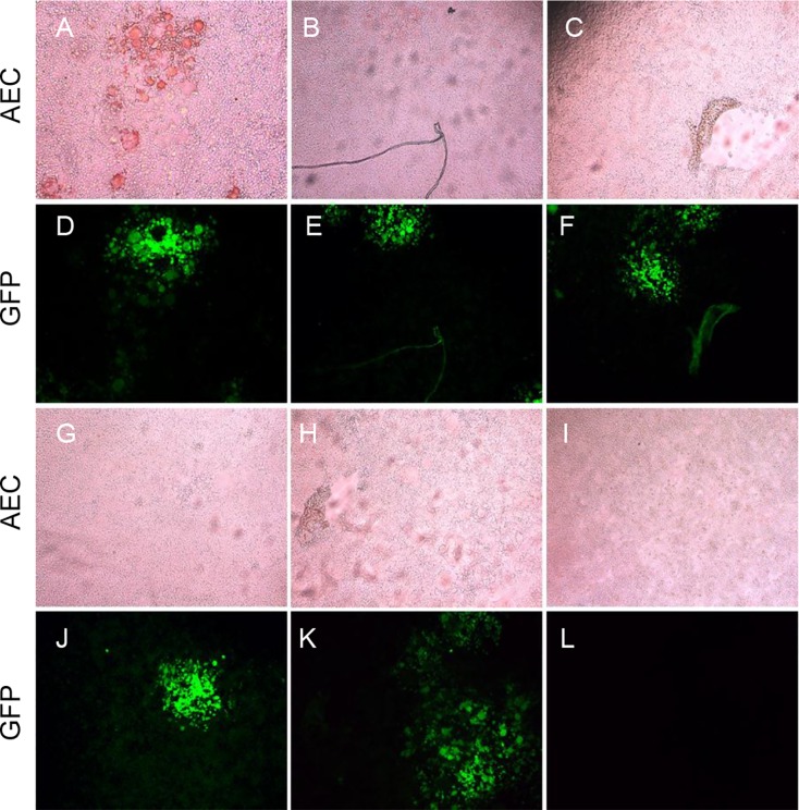
Fig 5. Specificity testing of the IPMA.
Panels A–C and G–H show the results of wells reacted with reference sera against PPRV, GPV, FMDV, BTV and Brucella. GFP expression is shown in panels D–F and J and K. Panel I shows the result of mock-infected BHK-SLAM cells. The results show that only wells reacted with PPRV sera were stained reddish-brown (panel A).

