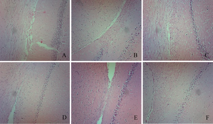
Fig 5. Pathology of focal cerebral ischemia/reperfusion rats (x100.0).
A, B, C, D, E, and F represent the sham group, the VT group, the rLj-RGD3 100.0 μg·kg-1 group, the rLj-RGD3 50.0 μg·kg-1 group, the Edaravone 1.5 mg·kg-1 group, and the Eptifibatide 100.0 μg·kg-1 group, respectively. Tissue pathology was observed using H&E staining. No edema, hyperemia, or other abnormal morphological characteristics were observed in the sham group. The cone cell array and cell boundary are clearly visible. In contrast, the ischemic group presented an edematous morphology with vacuolated architecture and pyknotic nuclei following H&E staining. In addition, the VT group rat brains showed obvious cone cell disarrangement, and their cell boundaries were not clear. Adverse effects were ameliorated in each treatment group.

