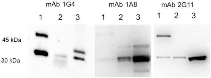
Fig 1. Reactivity of NheA-specific antibodies 1G4, 1A8 and 2G11 in a Western-blot assay.
Protein lysates applied were as follows: rNheA (1) supernatant of MHI 241 (2) supernatant of MHI 241 concentrated (3). The concentration was performed on Amicon® (Merck Millipore, Germany) Ultra centrifugal filters with a size exclusion of 30 kDa. The upper band in lane 1 represents rNheA with the 13 kDa thioredoxin tag still attached.

