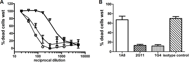
Fig 4. Results of the neutralization assays.
(A) Neutralization of toxic activity in supernatants from MHI 1507 by mAb 1A8 (open triangle), mAb 2G11 (black triangle) and mAb 1G4 (open dot). A high reciprocal dilution at the 50% toxic dose indicates a low or absent neutralizing capacity of the antibody applied. The neutralizing effect of 1G4 and 2G11 are similar while 1A8 and the isotype control (see S2 Fig) are not able to neutralize the cytotoxicity. (B) Consecutive neutralization of serially diluted antibody treated rNheA (applied to NheB/C (MHI 1761) primed cells. With mAb 1A8 or the isotype control the majority of the cells is dead. Error bars represent the SD of triplicates.

