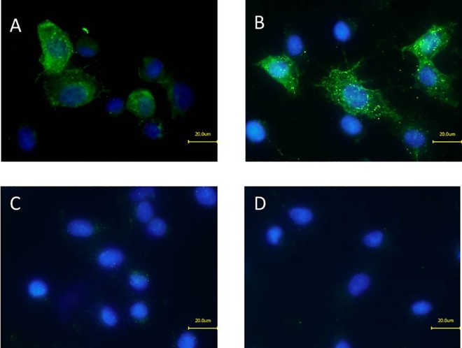
Fig 6. Immunofluorescence microscopy of NheA on Vero cells.
Staining of cell-bound NheA after treatment of Vero cells with B. cereus supernatants. (A) MHI 1507 NheB (100 ng ml-1 and approx. 10 ng ml-1 NheC) with mAb 1G4 neutralized NheA untreated Vero cells stained with primary and secondary antibody. (B) MHI 1761 (containing NheB at 100 ng ml-1 and approx. 10 ng ml-1 NheC) supplemented with neutralized rNheA (200 ng ml-1). (C) Stained Vero cells treated with rNheA only. (D) Untreated Vero cells stained with primary and secondary antibody. All slides were counterstained with DAPI.

