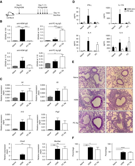
Figure 3.
Exposure to Pneumocystis antigen (PCAg) and house dust mite (HDM) generates lung pathology. (A) Schematic of treatment of mice with 10 μg of HDM and sonicated PCAg on Days 0, 7, 8, 9, 10, and 11. Naive (n = 9), HDM (n = 6), and PCAg (n = 10) mice were all killed at Day 14. Data from three individual experiments are shown. (B) PCAg and HDM exposure generated anti-PC and anti-HDM IgG and IgE responses, respectively. (C) PCAg and HDM increased expression of type II–related genes, as determined by quantitative reverse transcriptase–polymerase chain reaction. (D) Ex vivo cells isolated from whole lung were stimulated with either HDM or PCAg for 72 hours, and supernatants were analyzed for IL-4, IL-5, IL-17A, and IFN-γ. (E) Representative periodic acid–Schiff and hematoxylin and eosin staining of tissue from naive, HDM-treated, and PCAg-treated mice demonstrating increased mucus and perivascular inflammation in the HDM and PCAg groups. All images were taken at ×20 original magnification. (F) HDM and PCAg treatment increased the percentage of total airways affected by mucus (left) and the area of individual airway periodic acid–Schiff–positive staining (right) (*P < 0.05, **P < 0.01, ***P < 0.001, and ****P < 0.0001 by one-way analysis of variance with Tukey’s multiple comparisons). Hprt = hypoxanthine phosphoribosyltransferase; OD450 = optical density at 450 nm.

