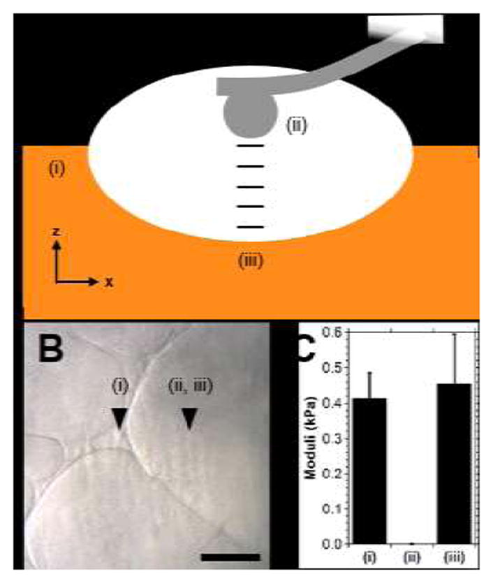
Fig. 6.

Examination of guest-host hydrogel heterogeneity via atomic force microscopy (AFM). (A) Schematic illustration of testing (side view), where moduli of the hydrogel (Day 3; 2.5 wt%) were determined at the top surface (i), in the pore center (ii, same z position as (i)) and at increments of approximately 10 μm until the bottom surface of the pore region (iii) was reached. (B) Bright field image, approximate location of testing indicated. Scale bar: 100 μm. (C) Indentation moduli at the locations tested. (mean ± SE; n ≥6; p = 0.77).
