. 2016 Oct 15;5:e17462. doi: 10.7554/eLife.17462

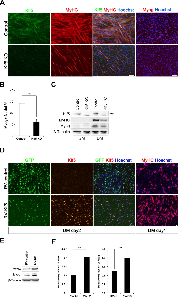
Figure 3. Klf5 is essential for muscle differentiation.

(A) Establishment of Klf5 knockout (KO) myoblasts using a CRISPR-Cas9 system. Klf5 KO C2C12 cells or Control cells were immunostained for MyHC and Klf5 or myogenin. Klf5 KO cells do not express Klf5 during muscle differentiation and exhibit severely reduced myotube formation. MyHC: myosin heavy chain, Myog: myogenin. Scale bar represents 100 µm. (B) Percentage of Myog-positive cells among total cells. Klf5 KO cells exhibited less Myog expression than Control. Data are means ± SEM. (***p<0.001) Representative data from at least three individual experiments are shown. (C) Western blots showing reduced MyHC and Myog expression in Klf5 KO C2C12 cells during differentiation. Representative data from at least three individual experiments are shown. (D) Klf5 KO cells were infected with a Retro-viral vector (RV-Klf5) harboring Klf5 or empty vector (RV-control), after which differentiation was induced for 2 or 4 days. The cells were then fixed and immunostained for Klf5 or MyHC. Impairment of myotube formation, as evidence from the loss of MyHC expression in Klf5 KO cells was rescued by exogenous Klf5 expression. Representative data from at least three individual experiments are shown. (E–F) Western blots revealing the reduction of MyHC and Myog expression in Klf5 KO C2C12 cells and its rescue by RV-Klf5. The expression levels were normalized to β-Tubulin (F). Data represent means ± SEM. (**p<0.01) Representative data from at least three individual experiments are shown.

DOI: http://dx.doi.org/10.7554/eLife.17462.006

Figure 3.

Figure 3—figure supplement 1. Klf5 knockdown impairs myoblast differentiation.

Figure 3—figure supplement 1.

(A) Klf5 expression during C2C12 differentiation. Bars depict means ± SEM. n = 3, biological replicates. GM: growth medium, DM: differentiation medium. (B) Induction of Klf5 expression during C2C12 differentiation was assessed immunostaining for MyHC and Klf5. Klf5 is highly expressed in the nuclei of differentiating myoblasts expressing MyHC and in myotubes. However, little Klf5 expression was seen in proliferating C2C12 cells in GM. Representative data from at least three individual experiments are shown. MyHC: Myosin heavy chain. Scale bar represents 100 µm. (C) Relative mRNA expression of Klf family members in C2C12 myotube on day 3 by QPCR. Bars depict means ± SEM. ****p<0.0001, ***p<0.001, **p<0.01, *p<0.05, N.S., not significant. n = 3, biological replicates. 
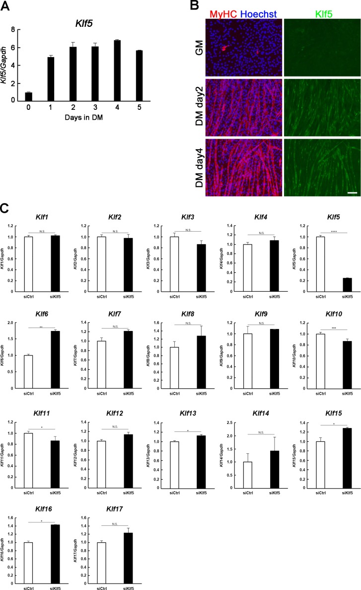
Figure 3—figure supplement 2. Establishment of Klf5 knockout C2C12 cells.

Figure 3—figure supplement 2.

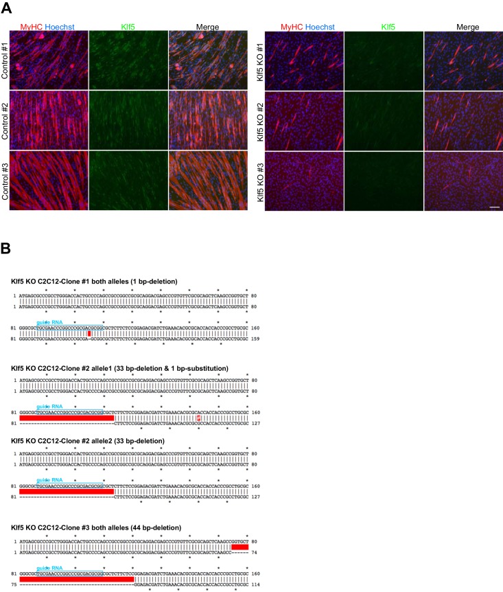
(A) C2C12 cells were transfected with pSpCas9-Klf5 or GFP-targeted control vector. After puromycin selection, the cells were cloned using the limiting dilution method. Three Klf5 knockout (KO) clones and three control C2C12 clones were obtained. Klf5 KO was confirmed by the absence of immunofluorescence. Compared to the control cells, all Klf5 KO cells exhibited defective myotube formation, as indicated by MyHC staining. Scale bar represents 100 µm. (B) Confirmation of mutation in the Klf5 gene locus. The position of guide RNA is indicated by the blue boxes. The red boxes indicate deleted sequences in the mutant allele, and the white # in the red box indicates the substituted nucleotide in the mutant allele. Clones #1 and #3 have a 1-bp deletion mutation in both alleles. The one allele of the clone #2 contains a 33-bp deletion, and the other allele contains a 33-bp deletion plus a 1-bp substitution mutation.
Figure 3—figure supplement 3. Phenotype of Klf5 knockdown cells.

Figure 3—figure supplement 3.

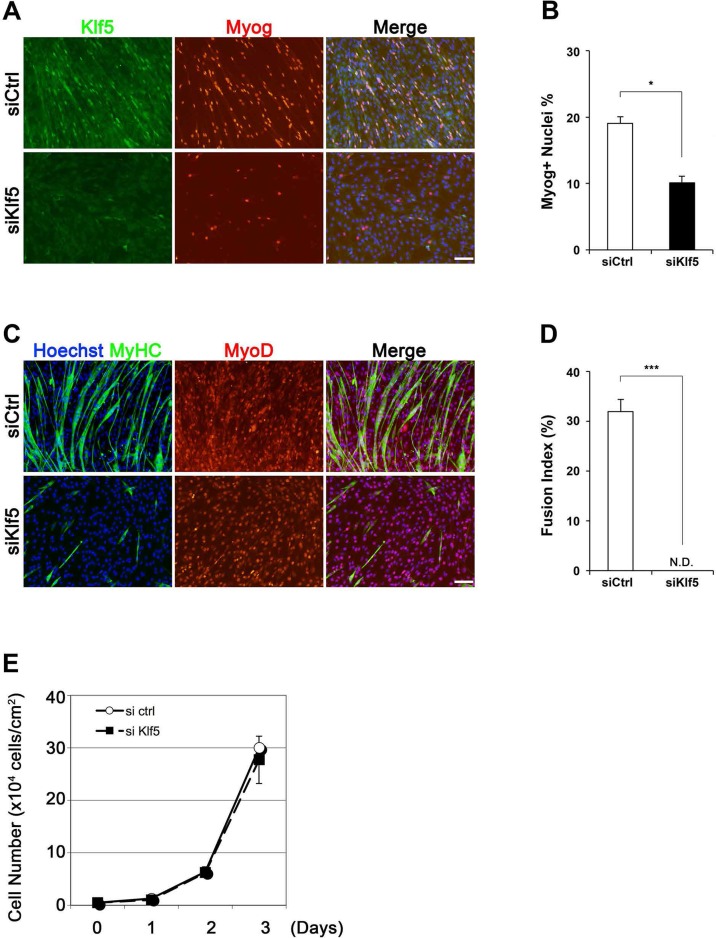
(AC) Klf5, myogenin (Myog), and MyHC expression in C2C12 cells after siRNA treatment. C2C12 cells were treated with either siKlf5 or siControl (siCtrl). After siRNA treatment in GM for 24hr, the medium was replaced with DM. After 2 days in DM, the cells were fixed and co-immunostained. Note the ratio of Myog-positive cell was significantly decreased by siKlf5 treatment (B). Data represent means ± SEM. (*p<0.05) Representative data from at least three individual experiments are shown. (D) Fusion index indicating fibers containing more than 3 nuclei. The fusion index was significantly reduced in cells transfected with siKlf5, though similar levels of MyoD positivity was detected. Data represent means ± SEM. (***p<0.001) Representative data from at least three individual experiments are shown. Scale bar represents 100 µm. N.D.: not detected. (E) Growth curve of siCtrl- and siKlf5-treated C2C12 cells in GM. Data represent means ± SEM. Representative data from four individual experiments are shown.