. 2016 Oct 15;5:e17462. doi: 10.7554/eLife.17462

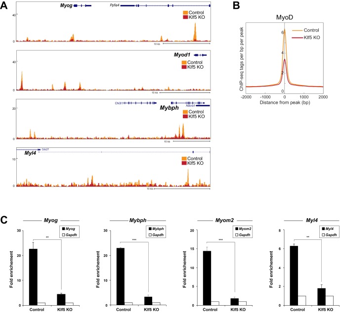
Figure 6. MyoD function is inhibited in Klf5-null C2C12 myoblasts.

(A) UCSC genome browser images illustrating normalized tag counts for MyoD at Myod1, Myog, Myl4 and Mybph gene loci in GFP-targeted control (orange) or Klf5-null (red) C2C12 myotubes differentiated for 3 days. (B) Distribution of MyoD tag densities in the vicinity of MyoD-bound enhancers in the GFP-targeted control or Klf5-null C2C12 myotubes differentiated for 3 days. (C) Comparison of MyoD recruitment at E-box containing enhancers of the Myog, Mybph, Myom2 and Myl4 gene loci in the GFP-targeted control and Klf5 null C2C12 myotubes differentiated for 3 days. Data represent means ± SEM. (**p<0.01, ***p<0.001, n = 3, biological replicates).

DOI: http://dx.doi.org/10.7554/eLife.17462.016

Figure 6.

Figure 6—figure supplement 1. Levels of MyoD and Mef2 protein were not altered by Klf5 deletion.
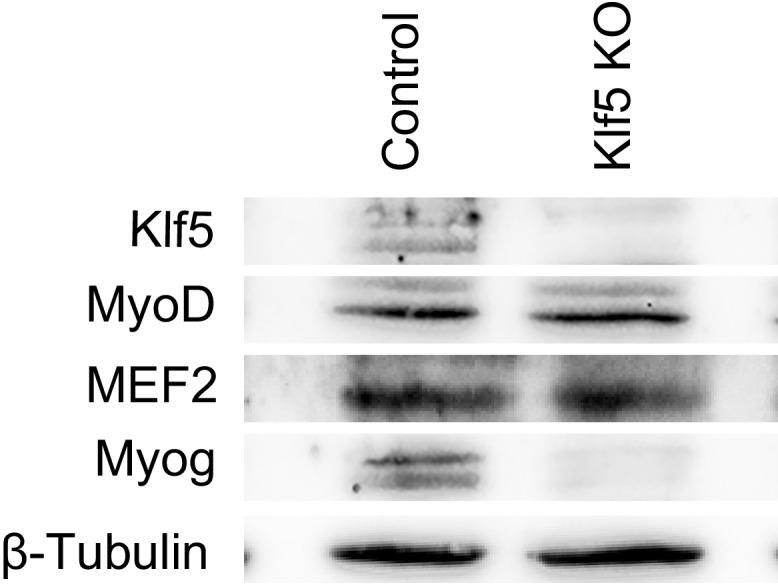
Figure 6—figure supplement 1.

Levels of MyoD, Mef2, Klf5 and Myog protein were analyzed by Western blotting using GFP-targeted control or Klf5 knockout myoblasts differentiated for 5 days. Representative data from four individual experiments are shown.
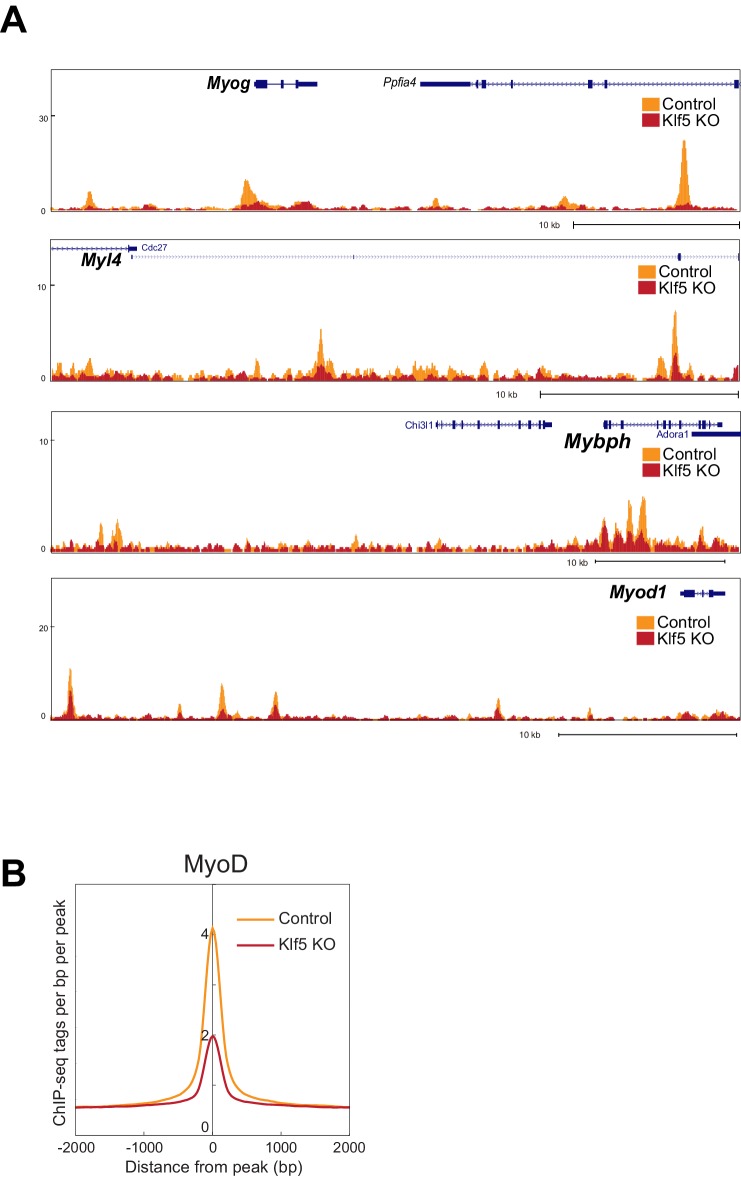
Figure 6—figure supplement 2. MyoD function is inhibited in Klf5-null C2C12 myoblasts -analysis in the another set of control and Klf5-null cells.

Figure 6—figure supplement 2.

(A) UCSC genome browser images illustrating normalized tag counts for MyoD at Myog, Myl4, Mybph and Myod1 gene loci in GFP-targeted control or Klf5-null C2C12 myotubes differentiated for 3 days. (B) Distribution of MyoD tag densities in the vicinity of MyoD-bound enhancers in GFP-targeted control or Klf5-null C2C12 myotubes differentiated for 3 days.