. 2016 Sep 13;5:e16761. doi: 10.7554/eLife.16761

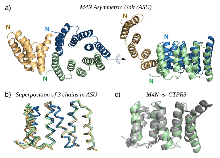
Figure 5. The X-ray structure of M4N.

(a) The three chains A, B and C in the asymmetric unit are colored green, blue and yellow, respectively. Chains A and B form a dimer. (b) Superposition of the three chains. Only Cα traces are shown for clarity. (c) Superposition of M4N (chain A, green) and the designed consensus TPR CTPR3 (PDB: 1na0, chain A, gray).

DOI: http://dx.doi.org/10.7554/eLife.16761.013

Figure 5.

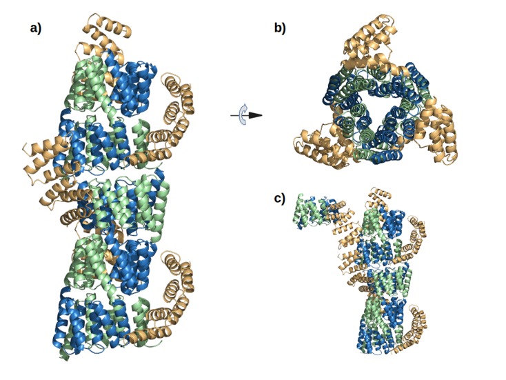
Figure 5—figure supplement 1. The interaction of M4N molecules in the crystal.

Figure 5—figure supplement 1.

(a) Five adjacent ASUs are depicted. Chain A (green) and B (blue) form a dimer, while chain C (yellow) packs its C-terminus to the N-termini of chains A and B. (b) Top view. (c) An additional ASU (top-left) is shown to demonstrate the packing of N-termini of chains C.
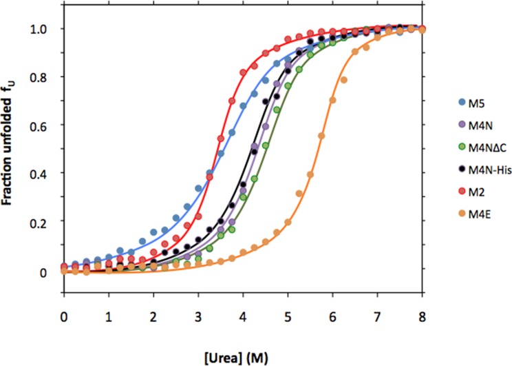
Figure 5—figure supplement 2. Urea denaturation of designed TPR repeats.

Figure 5—figure supplement 2.

Urea-induced equilibrium unfolding at 23°C was monitored by circular dichroism at 222 nm. Data were converted to the fraction of unfolded protein fU and fitted to a two-state model. The protein concentration was 15 µM. See Supplement file 1F for obtained parameters.
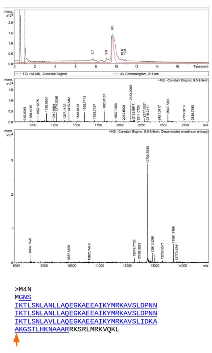
Figure 5—figure supplement 3. Mass spectrometry (MS) analysis of M4N.

Figure 5—figure supplement 3.

The M4N fragment with a mass of 12733.533 Da in MS is underlined and highlighted in blue (theoretical mass 12733.77 Da). The C-terminus of M4N as observed in the crystal structure is marked by a red arrow.