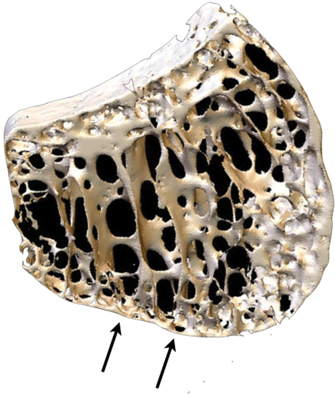
Fig. 1.

Microanatomy of the lunate. 3D micro-CT scan of the lunate demonstrating the thin proximal single layer of subchondral bone plate, which was measured to be 0.1-mm thick. There are spanning trabeculae principally on the radial aspect.13 3D, three-dimensional; CT, computed tomography. (Image courtesy of Dr. Gregory Bain.)
