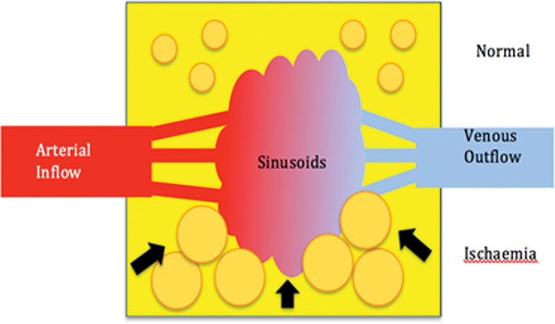
Fig. 8.

Compartment syndrome of bone. Diagrammatic representation of the normal vascularization of the lunate (superior half) with the normal fat cell and venous drainage. With ischemia (inferior half), there is interstitial edema and the marrow fat cells become swollen. This leads to tamponade of the sinusoids, thus decreasing the venous outflow. This further increases the intraosseous pressure, reduced arterial inflow, and produces necrosis. (Image courtesy of Dr. Gregory Bain.)
